KIMS General Surgery Department Successfully Operates A Rare Case of Pseudomyxoma Peritonei – “Jelly Belly”
Introduction

Pseudomyxoma Peritonei (PMP), nicknamed “Jelly Belly”, is a very rare disease characterised by the presence of jelly-like fluid called mucin in the abdominal cavity. What makes it so rare is that it occurs only in 1 to 4 per million per year, it is seen equally in men and women. First case was reported by a gynecologist, Dr. R. Werth, in 1884. Earlier the cause was unknown and the discussion regarding the etiology emerged very recently, in the first quarter of the 20th century. In 1 to 20% of the patients it is coincidently detected radio logically or at laparotomy or during hernia repair when mucus is found in the hernia sac. It is not a primary peritoneal disease. It’s a disseminated disease from a primary lesion of one of the intra peritoneal organs,
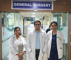
which being Appendix, Ovary, Colon, Stomach, Pancreas, and Urachus. From these organs the mucin breaks out and spreads into peritoneal cavity. The interval between the discovery of the primary tumour and the clinical diagnosis of PMP vary significantly (2 to 20 years) making it extremely difficult to diagnose in early stages. The complications associated with this condition are dreadful. As only 50% present with symptoms, the majority lies in the undetected zone, which can lead to serious complications like intestinal obstruction, severe ascites, menstruation problems & infertility. So, long term survival can be achieved by surgery alone. In cases where complete tumour removal is not feasible, maximum tumour debulking can still result in long term survival. PMP is challenging, complex but nevertheless the most rewarding peritoneal malignancy amenable to cure by CRS and HIPEC. Here in the Department of General Surgery, KIMS, we successfully performed this cytoreductive surgery in a rare case of pseudomyxoma peritonei, primary arising from the appendix.
Case Report
An elderly hypertensive lady was presented at OPD in February 2020, with distension and pain in abdomen for a period of 8 months. She also had history with a swelling of both lower limbs, vomiting, fever, cough, loss of appetite, weight and breathlessness. On examination of abdomen which was grossly distended, with dilated veins over it, everted umbilicus (Fig.1) with presence of generalised mild tenderness. Post radiological investigation, she was diagnosed with PMP.
The patient underwent Surgery on 12th of February 2020, Exploratory laparotomy (with Debulking procedure) under general anaesthesia, in which 12 litres of yellowish jelly was removed (Fig. 2). Parietal peritoneum involvement was seen and was accompanied by excision of the mass (Fig. 3), Appendectomy (Fig. 4), Type A Hysterectomy (Fig. 5), Omentectomy (Fig.6), Parietal peritonectomy (Fig. 7). Post histo-pathological assessment diagnosis of pseudomyxoma peritonei was made, with the primary organ involved being the Appendix (well differentiated mucinous adenocarcinoma).
Discussion
PMP is primarily described as a peritoneal disease looking like a myxoma but it’s neither a real myxoma nor a primary peritoneal disease. It’s a disseminated disease from a primary lesion of one of the intraperitoneal organs. It has two theories: 1) Theory of secretion (intra-peritoneal extravasations of mucus secretion). 2) Theory of collection (Intraperitoneal collection of gelatinous material and mucinous tumour cells). Gravity accumulates deposits in the pelvis. 50-80% patients will have symptoms reflecting the location of the primary or the metastatic tumour. 20-30% Female patients will be detected with ovarian mass and will be on evaluation for low abdominal pain or menstruation problem or infertility. 1-20% Patients are coincidentally detected on USG or CT scan or at laparotomy for any other reason when mucus is found in the abdomen. <1% Patients presenting with suspected bladder tumour or right femoral neuropathy. Earlier it was misunderstood that the incidence of PMP in females was more than males, which has been proven wrong in various articles, showing the incidence is equal in both sexes. In most female patients, it is commonly misunderstood that the primary arises from the ovary, likewise understood in our case, post surgery after histopathological examination, it was found that the primary arising from appendix, and the post op immunohistochemistry markers suggested the same.
Conclusion
What is the right approach? It varies from patient to patient, this patient was dealt with so carefully by KIMS’ elite team of surgeons for prolonged hours as mentioned earlier. But until post operative investigations showed that the primary was in the appendix, it was understood that the primary was in the ovary of this patient. Post operative period was uneventful. The patient was asked to start chemotherapy & follow up, however the patient did not undergo chemotherapy and the patient expired in the first week of April, i.e.; during Covid-19 lockdown.